| ePOSTER | ||
| Theme: eLearning |

|
 Send Email
| Abstract Title | ||
| A novel e-learning module: Ear and temporal bone 3D anatomy and surgical approaches | ||

|
Authors: |
Horace Cheng Nikolas Blevins Robert Jackler Sumit Agrawal |
Institutions: | Schulich School of Medicine and Dentistry, Western University, London, ON, Canada Department of Otolaryngology - Head and Neck Surgery, Stanford University, Stanford, CA, USA Department of Otolaryngology - Head and Neck Surgery, Western University , London, ON, Canada |
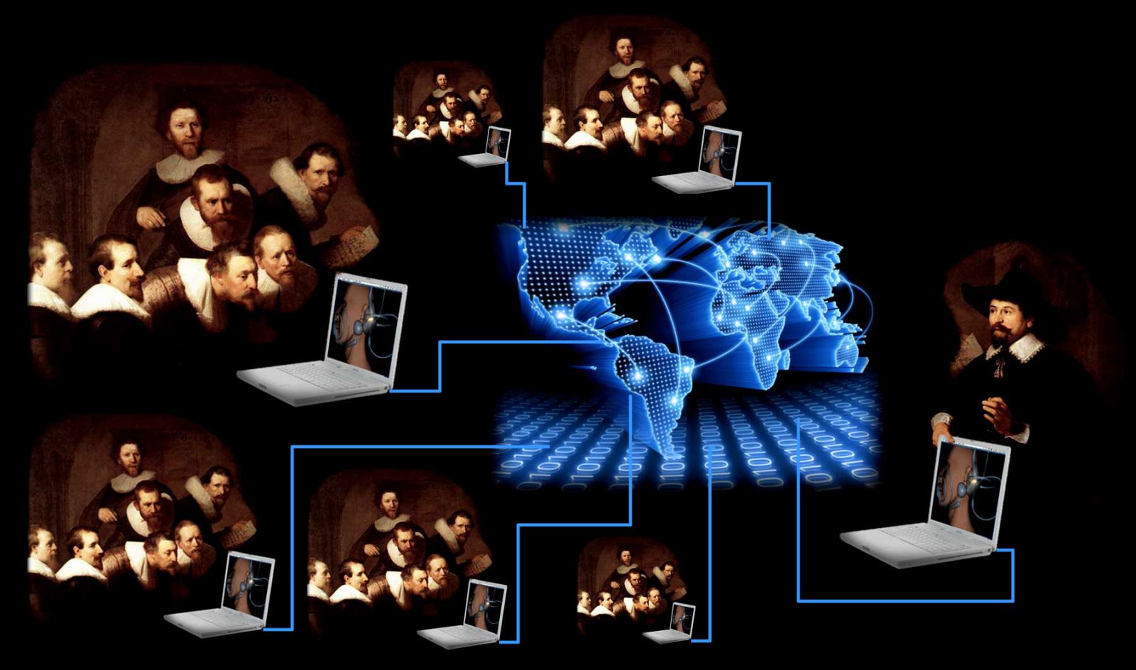
Rembrandt van Rijn (Dutch, 1606–1669). The Anatomy Lesson of Dr Nicolaes Tulp, 1632. Oil on canvas
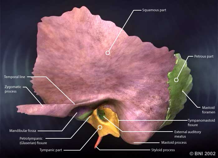
A good understanding of ear anatomy is important for medical students as it relates to the pathophysiology of otologic diseases. Likewise, a firm understanding of the anatomy and surgical approaches of the ear and temporal bone is essential to the training of otolaryngology residents. However, communicating the complex anatomy of these structures in traditional print medium has proved to be a challenge for medical educators and learners. Medical trainees of all levels have increasingly seek to enhance their learning experiences by the use of interactive computer based anatomy models. Attempts to create computer-generated, interactive, 3D models of the ear and temporal bone have been made but the widespread adoption of these useful learning aids have been hampered by the single-user delivery method and technology-compatibility issues.
This novel e- learning module is the result of collaboration between Western University and Stanford University. It is developed as a e-learning tool for medical students and otolaryngology residents. It uses a flashed-based platform specifically designed to be accessible online and compatible with all major browsers and tablet devices, thus overcoming the major limiting factors in delivery of material to learners. Software used in its development includes Adobe Captivate 6, Object2VR, Adobe Photoshop and Illustrator.
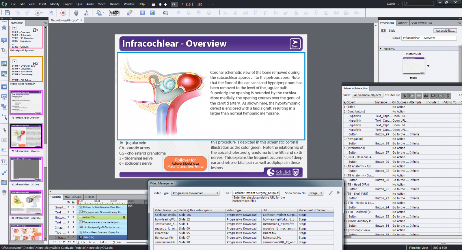
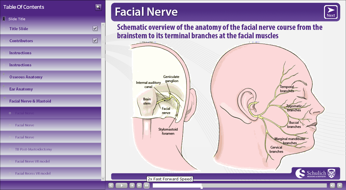
In addition to 2D anatomical and surgical contents from Dr. Jackler's 3D ATLAS OF SKULL BASE SURGERY & NEUROTOLOGY, this module also include models of Temporal Bone Dissector, interactive contents, and digitally-recorded HD surgical video.
This novel e-learning module is capable of delivering interactive world-class contents of ear and temporal bone anatomy directly to medical students and otolaryngology residents around the globe over the web.
Please take the opportunity to visit our website: http://tinyurl.com/neurotology

- 3D anatomy models created by Dr. Blevins
- 2D atlas and surgical contents provided by Dr. Jackler
- Supervision and guidance by Dr. Agrawal
- Funding and support by Otolaryngology - Head and Neck Surgery Department, Western University
- Funding and support by Undergraduate Medical Education Office, Schulich School of Medicine and Dentistry, Western University