


Theme
Simulation
INSTITUTION
University of Michigan Department of Neurosurgery
University of Michigan Department of Mechanical Engineering
University of Michigan Department of Medical Education

Purpose: In contrast to many other surgical fields, neurosurgery has yet to routinely incorporate simulators in resident education. We developed a high-fidelity lumbar spine simulator for teaching neurosurgical residents to perform minimally invasive techniques without risk of harm to patients in a low-pressure environment. Feedback from the first prototype guided refinements of the second prototype. We evaluated both prototypes for validity evidence relevant to test content and internal structure based on current Standards1.
Design
- Completely synthetic
- Bony model constructed from patient CT using 3D printer
-
Our model allows:
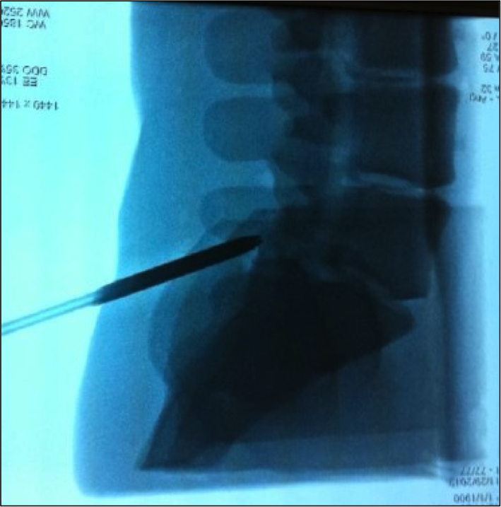
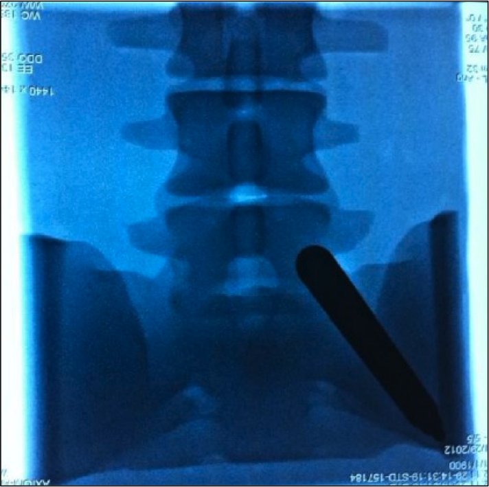
- Placement of tubular retractors with fluoroscopic guidance
- Drilling of bony structures
- Manipulation of thecal sac, nerve roots, disc fragments, etc.
- Filling of thecal sac with fluid for detection of CSF leak
Evaluation
Four neurosurgery residents and one attending neurosurgeon (n = 5) evaluated the first prototype. Eight residents and five attendings (n = 13) evaluated the second prototype. Evaluators incised, dissected, placed tubular retractors, and reviewed fluoroscopic images of the model. Participants then completed a 4-point rating scale, ranging from "Not at all realistic" [1} to "Highly realistic" [4] in:
- Physical attributes
- Realism of experience
- Ability to perform tasks
- Value and relevance (rated on a 5-point scale)
Fluoroscopic Images of simulator
The observed averages for the first and second prototypes, respectively, were:
- Physical attributes: 3.2 and 3.4
- Realism of experience: 3.1 and 3.6 (p = 0.047)
- Ability to perform tasks: 3.1 and 3.8
- Value: 4.6 and 4.9 (p = 0.005)
- Relevance: 4.8 and 4.7
The global ratings of the first and second forms of the prototypes were 2.6 and 3.2 respectively, indicating that the simulator "can be used as is for training, but could be improved slightly". Inter-rater reliability for the final prototype was high, ICC(2,13) = 0.81, 95% CI [0.61, 0.93], suggesting high participant rating agreement, offering validity evidence relevant to internal structure.
Computer rendering of MIS spine simulator
.png)
Final prototype ratings were high in all domains, indicating improved evidence in the content validity. Faculty and resident evaluation of the simulator indicate that it is a valuable training device with room for minor improvements.
Future Directions
- Incorporation of simulator into resident curriculum for training in MIS techniques, with evaluation of impact upon resident operative performance
-
Expansion of simulator application to include:
- Deformity
- Instrumentation
- Patient-specific anatomy
1American Educational Research Association, American Psychological Association, & National Council on Measurement in Education. (1999). Standards for educational and psychological testing. Washington, DC: American Educational Research
 Send Email