ePoster
Abstract Title | Investigating the effect of off-centre positioning of the heart in cardiac studies using the GE Discovery NM530c dedicated cardiac camera


Theme
Nuclear Medicine
INSTITUTION
CancerCare Manitoba, Winnipeg, MB., Canada

Background
- Myocardial perfusion imaging (MPI) is traditionally performed by using multi detector SPECT imaging.
- In recent years manufacturers started to create innovative designs of dedicated cardiac cameras. Most of the designs were aiming to simultaneously image the cardiac field of view by using the stationary detectors surrounding the region of interest.
- To enhance the performance of the new designs, the cadmium-zinc-telluride (CZT) solid state detectors, which have a superior spatial and energy resolution, were used.
- This significantly improves the image quality and reduces the acquisition time and/or patient radiation dose.
- The purpose of this study was to assess the impact of off-centre positioning of the heart in cardiac studies using the dedicated cardiac camera. In large patients (i.e. above ~140 kg) it is not possible to position the heart in the centre of the field of view. Concern was raised that this was affecting the appropriate visualization of tracer uptake and distribution within the myocardium.
Summary of Work
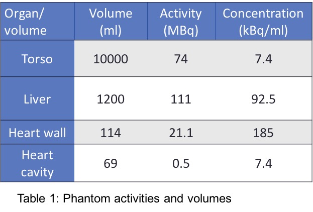
- A torso phantom which contains heart, lung and liver inserts was filled with water and radionuclide concentrations that roughly mimic clinical ratios between the above organs.
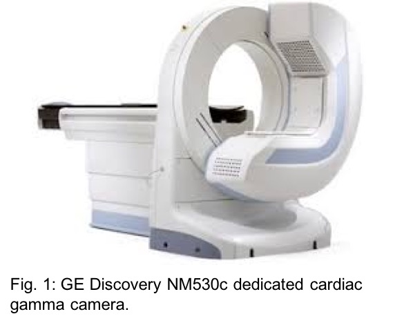
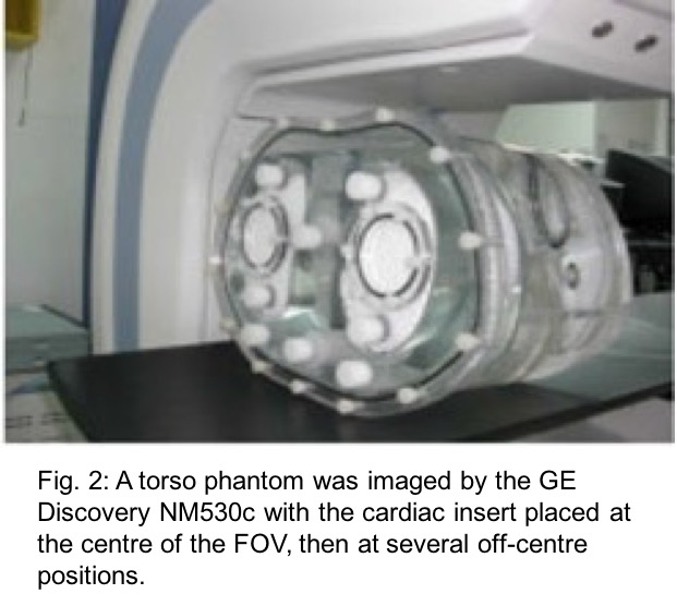
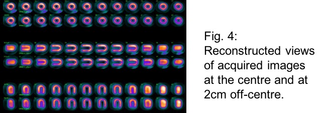
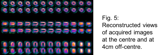
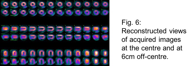
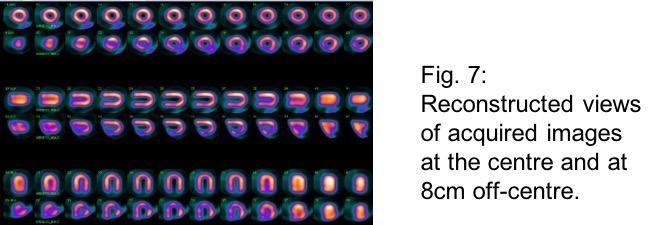
- The phantom was imaged by a dedicated cardiac camera (GE Discovery NM 530c) with the cardiac (heart) insert placed at the centre of the field of view, then imaged at several off-centre positions (1-8 cm off-centre within the FOV).
- Acquisition protocol: 2 days stress/rest, 5 minutes each image, heart insert was positioned in the centre then 1-8 cm off-centre within the FOV.
Summary of Results
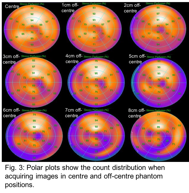
- This study shows that images acquired with the heart positioned a few centimeters off-centre of the FOV during cardiac imaging with the Discovery NM 530c camera can induce regions of count loss in the reconstructed images.
- In a clinical patient, these would be falsely interpreted as myocardial perfusion defects.
Conclusion
- For obese patients, performing myocardial perfusion SPECT, with the GE Discovery NM 530c CZT camera, may produce perfusion artifacts that are variable in extent and location (i.e. not predictable) as the patient's heart can’t be positioned within the centre of the FOV.
- Thus, if the optimal patient positioning can’t be achieved with the CZT camera, it is important to consider alternative solution including schedule the patient on a conventional SPECT camera for cardiac imaging.
 Send Email