


Theme
Diagnostic & Interventional Radiology
Title
Case Report of Hydatid Cyst in the Pulmonary Artery Uncommon presentation: CT and MRI findings
Background
Hydatid cyst can be found in any organ as primary echinococcosis or because of spread from other organs as secondary echinococcosis. In adult, the liver and lungs are the most common locations, pulmonary artery involvement by hydatid cyst is rare.
Summary of Work
Clinical case: We report a rare case of an 86 years old female with a history of hepatic hydatid cyst since 2012, presenting with pulmonary artery hydatid cyst located in the right pulmonary artery, and complains of chronic productive cough, yellow-greenish sputum, and dyspnea. She lives in the rural area. Chest x-ray, computed tomography, and magnetic resonance imaging were done. Chest x-ray showed a mass -like opacity in the right lower zone, right paracardial area. CT showed pulmonary artery with multiseptated hydatid cysts. MRI confirmed the presence of cystic lesion within the right pulmonary trunk extending to the right lower lobe pulmonary artery showing low signal intensity on T1 and high signal intensity on T2 with septation. The patient refused surgery and was discharged on oral Albendazole 400 mg twice daily and oral Praziquantel 1800 mg twice weekly. After ten months, the patent had CT and MRI which showed mild regression of the size of both right main pulmonary artery and 2 left sided hydatid cysts. Praziquantel was stopped and now she is only on oral Albendazole 400 mg twice daily.
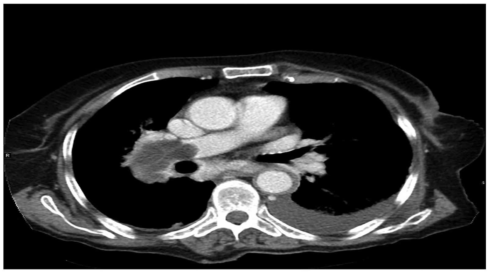
Figure 1: CT axial image shows pulmonary artery with multiseptated hydatid cysts, seen in the right main pulmonary artery and the lower lobar branches of the right pulmonary artery.
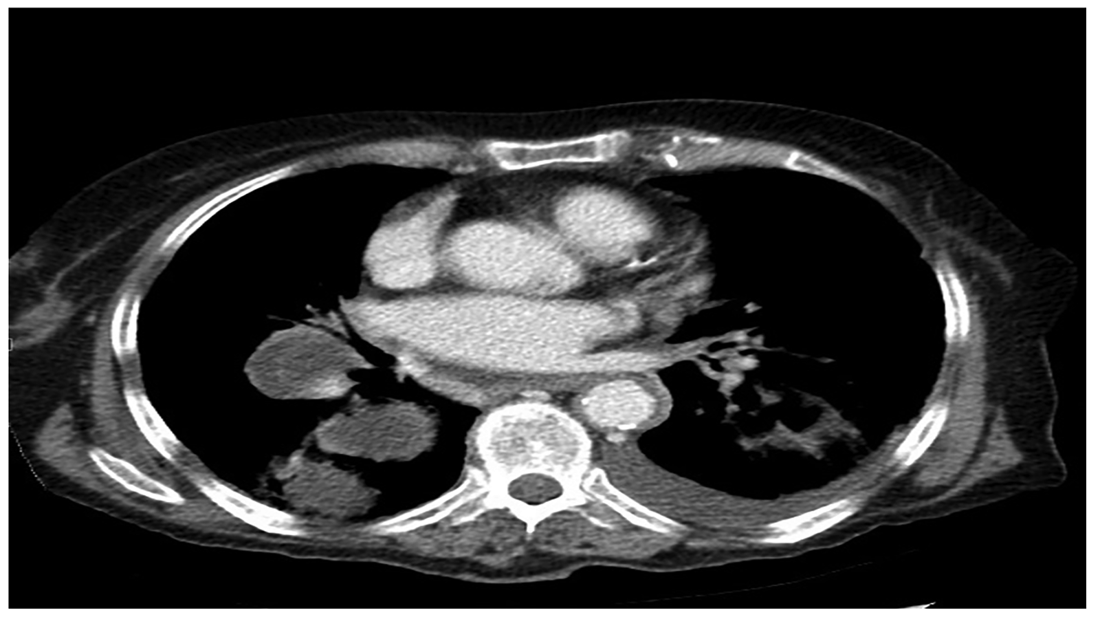
Figure 2: CT axial image shows pulmonary artery with multiseptated hydatid cysts, seen in the right main pulmonary artery and the lower lobar branches of the right pulmonary artery, along with other parenchymal cysts
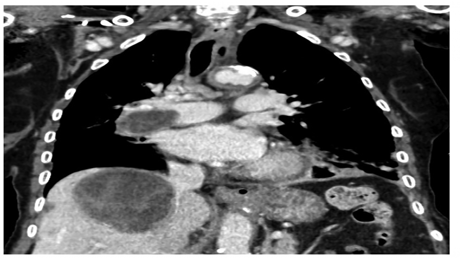
Figure 3: CT coronal images show right main pulmonary artery hydatid cysts along with a cyst of the right hepatic lobe.
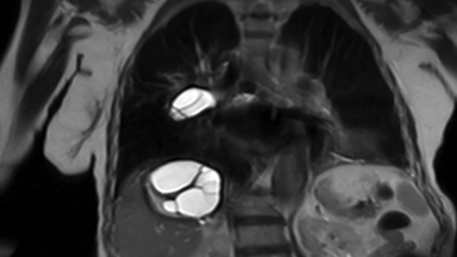
Figure 4: A coronal T2 weighted image shows a high signal intensity cystic lesion within the right main pulmonary artery with multiseptated high-intensity cystic lesion on the right hepatic lobe.
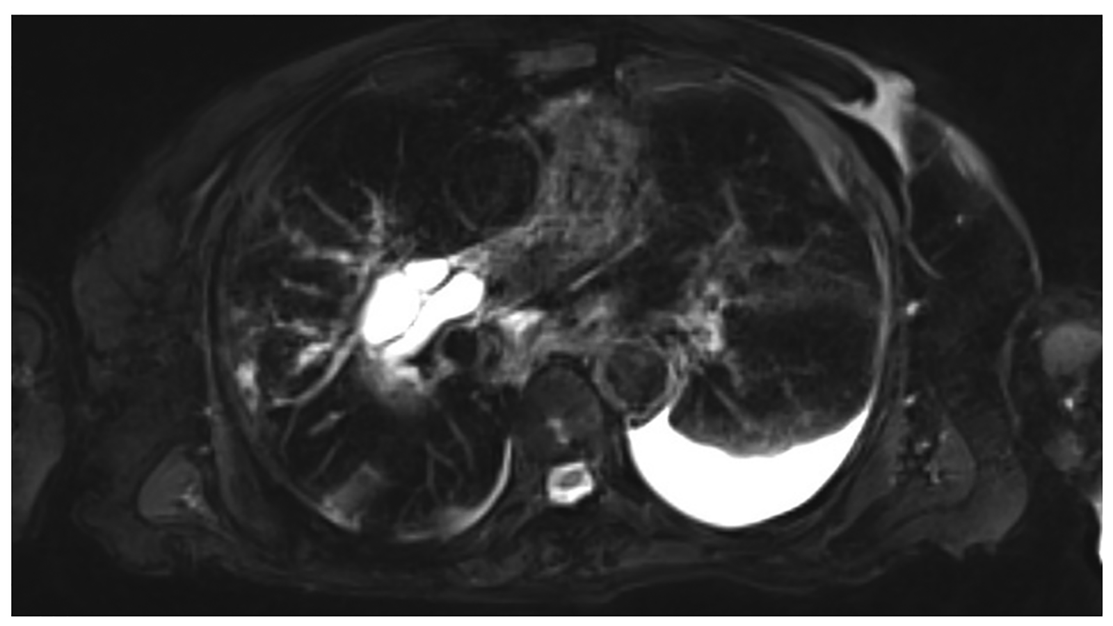
Figure 5: Axial T2 weighted image shows hydatid cyst in the right main pulmonary artery and small pleural effusions on the left side.
Conclusion
Recognizing the possibility of the presence of hydatid cyst in pulmonary artery is important. Also, knowing the features on CT and MRI may aid in the differential diagnosis.