


Theme
Radiation Protection & Radiobiolgy
INSTITUTION
King Saud University
King Faisal Specialist Hospital and Research Center

The quality assurance (QA) program in diagnostic X-ray facilities involves techniques that are routinely necessary to ensure that X-ray Machines are working accurately and providing a high diagnosis service with maintaining high imaging quality at minimum risk to patients and workers. The most important aspect of a QA program is the measurement of the dose received by individuals during examinations and checking the X-ray beam parameters such as the first Half Value Layer (HVL) which defined as the amount of aluminum thickness that is required to attenuate the beam output to one half of its initial value.
Our work was aiming at enhancing the reference X-ray beam qualities (RQR) through optimizing HVL measurements to establish the best measurement and calibration capabilities for diagnostic radiology at the Secondary Standard Dosimetry Laboratories (SSDL) belonging to King Faisal Specialist Hospital and Research Center (KFSHRC) in Riyadh (Saudi Arabia).
Establishment of the RQR
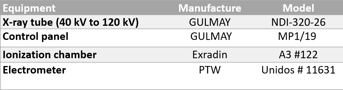
The equipment characteristics used to perform this work are presented in Table(1). The RQR defined as a measure of the penetrative power of the radiation beam emerging from the X-ray machine, usually characterized by a statement of the tube potential and the first HVL.
Table 1. Equipment used at SSDL in the HVL measurements.
To establish the RQR qualities, the attenuation curve was measured using different Aluminum attenuators.
As a first step, measurement was performed without any additional filtration. For each RQR quality, the corresponding voltage (40 to 150 kV) was selected and the tube current was fixed to 15 mA.
The next step is the determination of the additional filtration necessary to achieve the desired RQR qualities, the method recommended by IAEA TRS No. 457 and the IEC 61267 was followed.
The final step is, to add the additional filtration determined above for each RQR quality and to determine the corresponding HVLs, the Aluminum attenuators were added gradually behind the collimator, Fig (1) illustrates the set-up used for HVLs measurements.
The first HVL is determined by fitting the attenuation curve using a commercially available curve fitting software (Tablecurve).
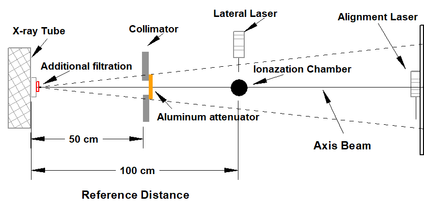
Fig 1. Experimental setup used for the determination of HVLs
Optimization of HVL measurements
To determine the optimal additional filtration values giving minimal deviation to the IEC HVL values, for each beam quality, we have applied the trial and error measurements. If the ratio of the measured HVL (calculated homogeneity factor h) is below 0.485 (deviation less than -3%) then the additional filtration needs to be increased and vice versa. To allow interpolate for the optimum addition filtration, at least three attenuation curves were measured for three additional filtrations around the nominal value given by the IEC standard.
Air kerma measurements
The air kerma rate was measured for the optimum HVLs that were identified previously.
At this work, the reference chamber Exradian A3 (see Fig 2 ) was positioned at 100 cm from the focal point of the X-ray tube and connected to an electrometer, the tube current was fixed to 15 mA. Fig (3) illustrates the set-up used for the air kerma rate measurements.
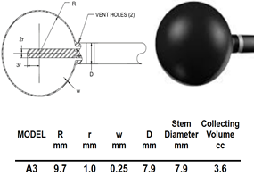
Fig 2. Characteristics of the Exradin reference spherical chamber
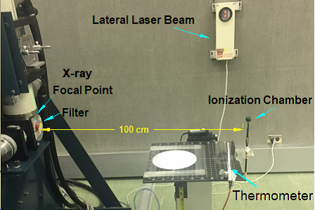
Fig 3. Experimental set-up used for the air kerma rate measurements
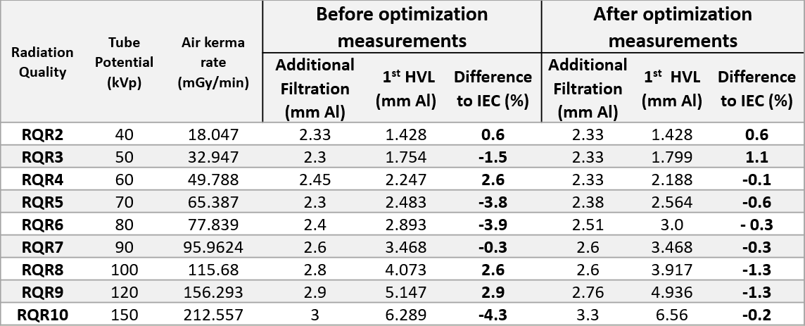
Fig (4) shows the results of the attenuation curves used for HVL measurements. The overall optimization process is summarized in Table (2) giving for each beam quality the measured first HVLs before and after optimization. As can be seen, a net improvement in the difference to the IEC 61267 standard is obtained.
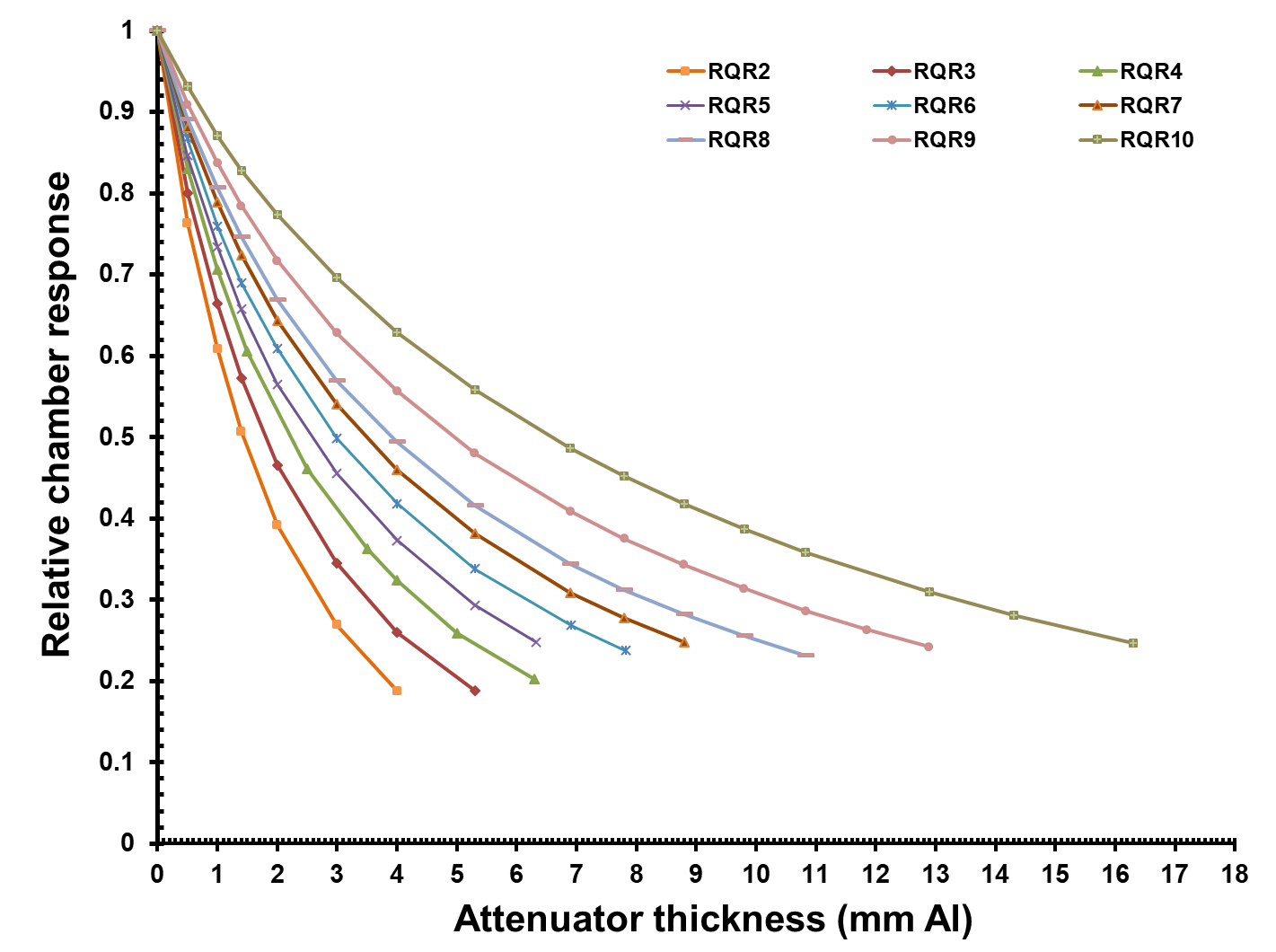
Fig 4. Attenuation curve used for HVL measurement for all RQR qualities
Table 2. First HVL measurements at SSDL before and after optimization processes
For air kerma measurements, a linear fit of the air kerma rate versus the 1st HVL was obtained with a correlation coefficient R2 equal to 0.999 showing a perfect linearity between the two quantities. A linear equation was derived Fig (5).
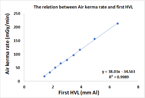
Fig 5. Variation of air kerma rate versus first HVL
The optimum additional filtrations (from 2.33 to 3.3 mm Al), were measured accurately at SSDL. The corresponding first HVLs are well within the IEC accepted criteria of +/- 3 %.
The best measurement capabilities for the diagnostic radiology RQR beam qualities, given in terms of air kerma rates, are from 18.05 to 212.56 (mGy/min).
Wish to thank the administration of KFSHRC represented by Dr. Sultan T. Alsedairi, Dr. Belal Moftah, and Mr. Fareed Mayhoub, for allowing me to undertake my MSc thesis at KFSHRC and utilizing the laboratory facilities.
Special thanks to my supervisor Prof. Dr. Mehenna Arib and Mr. Omar Noor, for their clinical/theoretical knowledge as a medical physicist were very obvious when helping me perform the experimental measurement, Thank you.
Also, I would like to thank my supervisor Prof. Dr. Mohamed Abdelhalim for his help and support to make this work possible
1. Faison, C. and Brickenkamp, C., 2004, Calibration laboratories, 1st ed, Calibration Laboratories Technical Guide for Ionizing Radiation Measurements, National Institute of Standards and Technology, US.
2. IAEA, 2007, Dosimetry in Diagnostic Radiology: An International Code of Practice, Technical Reports Series No. 457, IAEA, Vienna.
3. IEC, 2006, Medical Diagnostic X-ray Equipment-Radiation Conditions for Use in the Determination of Characteristics, IEC-61267.
4. Ma, C.M., Coffey, C.W., DeWerd, L.A., Liu, C., Nath, R., Seltzer, S.M. and Seuntjens, J.P., 2001, AAPM protocol for 40–300 kV X‐ray beam dosimetry in radiotherapy and radiobiology, Medical physics, 28(6), pp.868-893.
5. Périard MA, Chaloner P, 1996, Diagnostic X-Ray Imaging Quality Assurance: An Overview, Consumer and Clinical Radiation Hazards Division Radiation Protection Bureau, Environmental Health Directorate Health Protection Branch, Health Canada.
6. WHO, 1982, Quality Assurance in Diagnostic Radiology, Geneva.
 Send Email