
| Theme: 9II Virtual Patients/Social networking | |||
 |
||||||
| Augmented Reality E-Learning for Medical Education |
 |
|||||
|
||||||

The study of pathology is inherently a part of medical education. The Gordon Museum of Pathology at King’s College London (KCL) houses the largest medical museum in the UK with text descriptions of over 8,000 specimens to support the learning of over 10,000 healthcare students and staff each year. But text descriptions alone may lead to misinterpretation and can be time consuming. e-Learning is a new mode of teaching which enables interactive learning, combined with multi-sensory inputs, e.g. visual and audio [1-3].
Augmented reality (AR) is a technology that allows virtual content, such as images and annotations, to be overlaid onto a live screen capture of physical objects in realtime, delivered an a mobile computer device (Figure 1). AR content was developed for the Gordon Museum using the Layar software platform, (Layar BV, Amsterdam, The Netherlands). Content was developed for six specimens, three of which showed different stages and complications of myocardial infarction (MI), and other other three showing different types of cerebrovascular accident (CVA).
This study investigated the impact on learning of medical students using augmented reality (AR) enhanced specimens, compared to the text-only descriptions available at the museum.

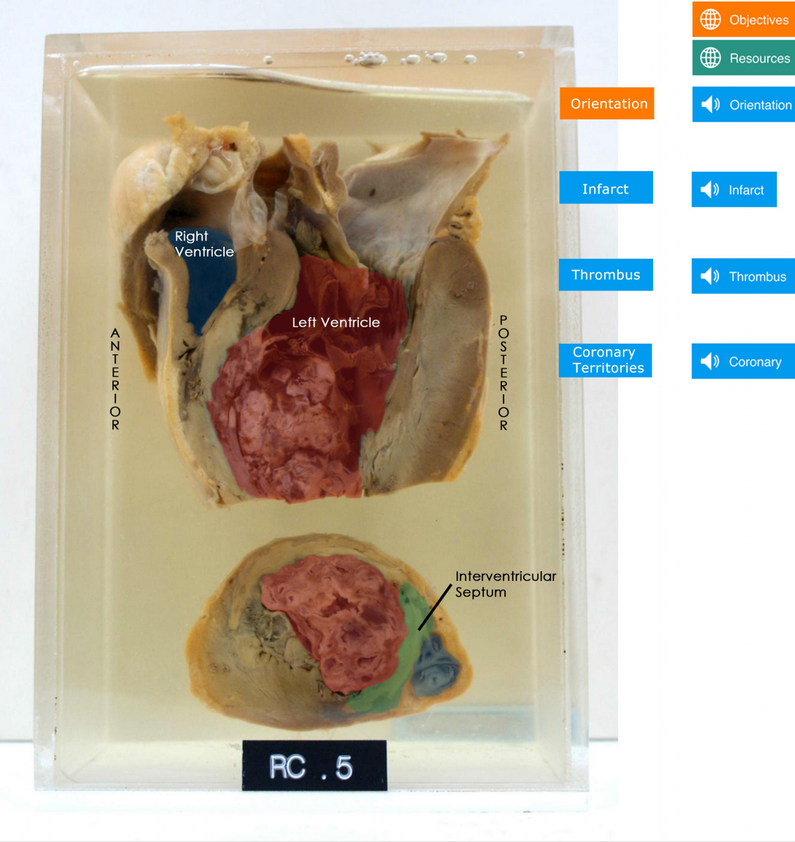
Figure 1) left): A photograph of a pathological specimen from the Gordon Museum. It shows sagittal (top) and axial (bottom) cross sections of a heart that has undergone a myocardial infarction. right): One of the overlays that appears when scanning the specimen in a) using the AR application. Interactive buttons allow the user to view different image and annotations overlays while listen to an audio description of the specimen.
Twenty KCL medical students were block randomised to either an intervention or control group. The intervention group used AR-enhanced specimens, created with Layar (Layar B.V., the Netherlands) and delivered via a tablet device. The control group used specimens with textonly descriptions as normally provided by the Museum. All participants completed objective assessments of learning outcomes before and after two timed sessions. There were three specimens in each session with the first concerning myocardial infarction (MI) and the other cerebrovascular accidents (CVA). Feedback was used for subjective assessment.
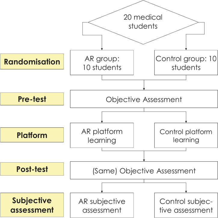
Figure 2: Outline of the study design.
This process was repeated twice, for the three MI specimens and for the three CVA specimens. This objective assessment covered the visual recognition of anatomy, pathology and the understanding of pathophysiological processes. These tests were structured to simulate OSCEs (Objective Structured Clinical Examinations) which are part of the KCL School of Medicine curriculum.
Objective Results
Students had greater improvement using the AR platform when visualising anatomy and pathology. This was not shown for pathophysiology for which the difference was statistically insignificant (Figure 3).
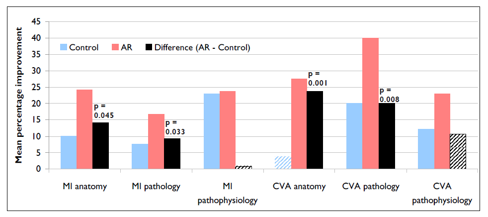
Figure 3: The mean improvement of test scores is displayed expressed as a percentage of the total available marks, where shaded columns represent statistically insignificant improvement (p>0.05). The benefit of AR over the control is shown as the difference; where significant (p<0.05), p-values have been displayed.
Subjective Results
The results of the rating scales correlate well with the objective results, showing AR to be preferred for visualising anatomy and pathology (Figure 4). Comments sections demonstrate that AR was overall the preferred platform (Table 1).
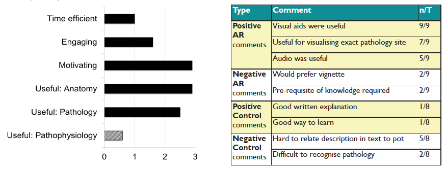
left) Figure 4: The difference between AR and control mean ratings are displayed, for each statement (calculated as AR mean - control mean). Shaded columns represent statistically insignificant differences (p>0.05). right) Table 1: The most common comments are displayed for both groups. T is the number of candidates who completed the questionnaire from that group, and n is the number that made that comment. Similar comments were categorised under a common theme.
The results of this pilot study show that AR was superior to traditional text descriptions when learning using anatomical specimens, particularly for visual recognition of anatomy (MI +13.3%, CVA +23.8%) and pathology (MI +9.2%, CVA +20.0%). However, this was not true for the understanding of pathophysiology, where no statistical significance was found. As a whole, students preferred the AR experience (68% positive comments) to traditional methods (85% negative comments).
AR is a powerful visual tool and may have an important role in enhancing learning for medical students.
Funding from the College Teaching Fund, King's College London
Greg and Alex, Initiator SSC students
Ian Johns and Martin Holland from Information Technology
Medical students volunteers who took part in our study
1. Klatt, E. C. and S. E. Dennis, 1998. Web-based pathology education. Arch Pathol Lab Med. 122:475–479
2. Dick F, Leaven T, Dillman D, Torner R, Finken L., 1998. Core morphological concepts of disease for second year medical students. Hum Pathol;29:1017–20
3. Reid WA, Harvey J, Watson GR, Luqmani R, Harkin PJ, Arends MJ. Medical student appraisal of interactive computer-assisted learning programs embedded in a general pathology course. J Pathol. 2000 Aug;191(4):462-5
 Send Email